Starfish Dissection Lab
INTRODUCTION
The phylum Echinodermata includes
starfishes or sea stars, brittle stars, sea urchins, sea lilies, and sea
cucumbers.
All but the last have an internal skeleton composed of
calcium carbonate material and hard external spines or plates.
They are fixed or slow-moving inhabitants of the
sea, from the high-tide zone to considerable depths.
They are often abundant, but no species form
specific colonies.
Species that live in shallow water are easily
collected by had at low tide and dredging captures deeper ones.
Those with skeletons are easily prepared merely by
drying, but specimens for dissection are preserved in methyl alcohol.
(Not formaldehyde!)
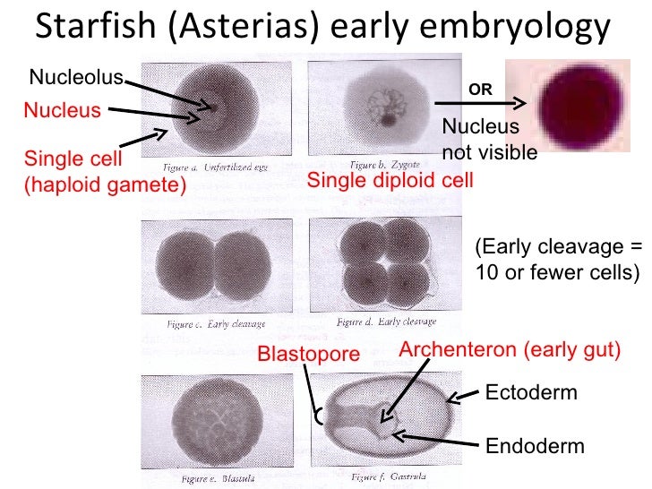
Eggs of starfish and sea urchins can readily be
obtained in quantity and fertilized as needed; hence, they serve for study in
embryonic development and in many experimental researches on animal eggs.
The common species of starfish used
for classroom observation and discussion is
Asterias forbesi.
Below is the complete taxonomy of the common
starfish.
KINGDOM:
Animalia
PHYLUM:
Echinodermata
CLASS:
Asteroidea
ORDER:
Forcipulatida
FAMILY:
Asteriidae
GENUS:
Asterias
SPECIES:
Asterias
forbesi
or
Asterias rubens (Asterias
forbesi…however is the more widely accepted species name)
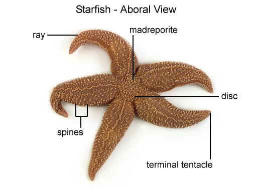
PART ONE: THE
EXTERNAL ANATOMY
1.
Place the starfish on your
dissection tray with its aboral surface facing upward.
2.
Using your diagram sheets locate
the following structures, and note their functions:
¨
Arms or Rays—these are the
five extensions that you see projecting from the middle of the starfish.
These are highly regenerative and are replaced
easily when damaged.
You might even see one of our starfish that has a
ray that is significantly smaller than the rest of them.
This is because the ray had been damaged or lost and
it has regenerated a new one.
The two rays which are closest to the madreporite
are known as Bivium.
¨
Central Disk—this is the
middle area of the starfish from which the rays extend.
It is often poorly defined and difficult to locate
the perimeters, but on some you may be able to distinguish a pentagon shaped
area.
¨
Aboral Surface—the aboral
surface is the surface that does not contain the mouth.
¨
Madreporite—this is a small,
white, circular area that is located in the central disk area.
It is usually off-center and is sometimes called the
sieve plate.
It is used by the starfish to take in water to fill
its water-vascular system.
If you scratch it with your probe you will notice
that it is rather hard and feels like stone.
¨
Anus—the anus is rather
minute and difficult to see but it is also located on the central disk.
Wastes are excreted through this opening to the
outside of the starfish.
¨
Spines—the entire aboral
surface is covered with many short, rough, limy spines
¨
Eyespot—the eyespot is
located at the distal end of each ray.
It is a collection of photosensitive cells which the
starfish uses to detect light or absence of light.
3.
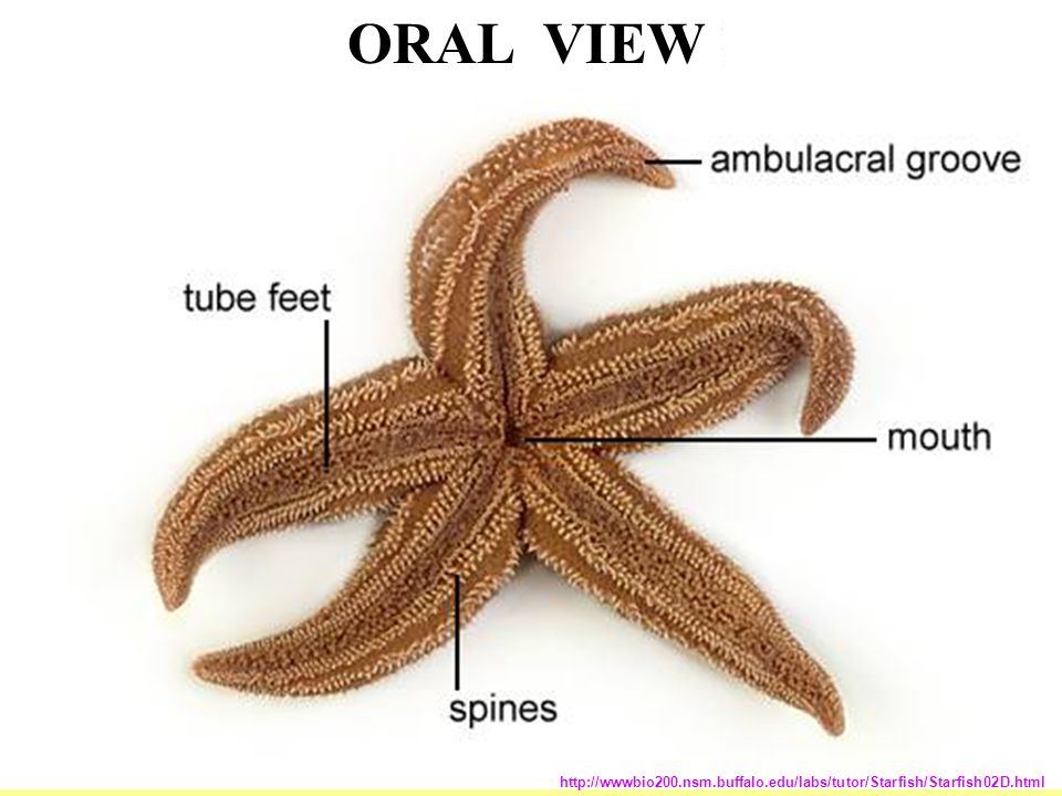
Now flip your starfish over so you
can view the oral surface.
Use your diagram sheets to identify the following
structures:
¨
Ambulacral Groove—this is
where the tube feet are located.
They are found along each ray.
¨
Ambulacral Spines—these are
slender rods located on the margins of the ambulacral grooves.
¨
Tube Feet—soft, slender, with
expanded tips.
There are two or more rows in each ambulacral
groove.
¨
Mouth—opening in the middle
directly beneath the central disk where all the arms connect.
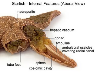
PART TWO: THE
INTERNAL ANATOMY
1.
Use the scissors to cut off the
extreme tip of each arm of the bivium.
Then cut along the sides of these two arms.
Use care not to injure any internal organs.
2.
In turn, lift and carefully remove
the aboral surface of each arm, loosening the delicate mesentaries beneath by
which the soft organs are attached.
Also, cut around the central disk to expose the
stomach underneath.
3.
Use your diagram sheets to identify
the following structures:
¨
Coelom—space containing the
internal organs; lined with thin ciliated peritoneum
¨
Stomach—sack-like structure
found underneath the central disk.
¨
Retractor Muscles—small,
sinewy structures that connect to the stomach.
These are used to pull the stomach back into its
mouth once the starfish is done feeding.
¨
Hepatic caeca—long,
greenish organs found in each ray.
Has many finger-like lobes.
These organs are used for secreting digestive juices
and enzymes needed for feeding.
¨
Gonads—small, bi-lobed
structures found below the hepatic caeca in each arm.
May be very small in some specimens due to the fact
that the starfish may not be sexually mature yet.
4.
In order to determine the sex of
your starfish, you must examine a small portion of the gonad with the
microscope.
5.
Make a mounted slide by taking a
SMALL PORTION of the gonad and placing it on the microscope slide.
Cover this with a cover slip and observe under the
low power objective.
6.
If your starfish is a female you
will see the eggs.
This resembles small circular-looking objects.
7.
If your starfish is a male you will
see the sperm.
Instead of seeing circular objects you will see
something that resembles sand.
These are the sperm cells.
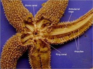
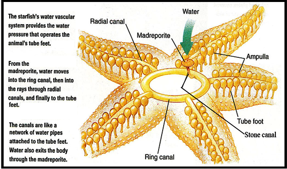
PART THREE:
THE WATER VASCULAR SYSTEM
1.
Remove the stomach from the central
disk area.
2.
You will now be able to see the
calcite skeletal system of the starfish and also the parts of its water vascular
system.
Use you diagram pages to locate the following structures:
¨
Ring
Canal—hard,
calcium-based, ring-like structure around the mouth region.
¨
Tiedemann bodies—nine, small
swellings in the ring canal.
¨
Ampullae—many, small,
spherical structures in the floor of the coelom.
These connect to the tube feet.
¨
Tube feet—tiny
extensions below the ampullae that fill the ambulacral groove, used for
locomotion
3.
Dispose of your starfish in the
garbage and clean up your trays and utensils.
4.
Complete the conclusion questions
about the starfish.
Click
HERE for the Starfish Dissection Lab
Companion